 Amyloodinium
sp.

 Amyloodinium
sp.
Amyloodinium sp. is a microscopic, single cell dinoflagellate which is parasitic on the skin and gills of tilapia and other fish in brackish to salt water environments. The organism, or at least this group of organisms, has a worldwide distribution in tropical to subtropical waters. Very similar organisms occur as parasites of freshwater fish. However, infections of tilapia in the islands have yet to be recognized in tilapia cultured or held in freshwater.
The trophonts of Amyloodinium sp. attach to the skin or gills of the tilapia. Once mature the trophonts form a cyst stage that gives rise to 200 or more dinospores. The dinospores are motile and are the stage of the parasite that attack new hosts. The dinospores remain infective for at least 13 days after release from the cyst. Dinospores that do not contact a fish during this time will die.
If conditions are proper, Amyloodinium sp. can multiply rapidly (e.g. multiple cysts release many infective dinospores) and when present in large numbers, particularly on the gills, the infection will result in high mortality of tilapia. Field observations in Hawaii have shown that tilapia transferred from freshwater to brackish or full strength seawater are particularly prone to heavy, serious infections by Amyloodinium sp. These fish are thought to have no natural immunity because in freshwater there was no prior exposure to the organism.
Amyloodinium sp. occurs commonly on wild tilapia in brackish water as an inapparent or subclinical infection. These fish are typically resistant to overt disease from this organism unless the tilapia are severely stressed or crowding results in extremely heavy infections. Tilapia from natural brackish waters should always be considered as carriers for Amyloodinium sp. and other ectoparasites. Never introduce these fish directly into culture systems without prior quarantine, treatment or a period of rearing in freshwater.
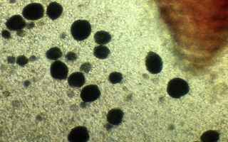
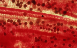
Amyloodinium sp. is non-motile, spherical to pear shaped and attached by a short stalk to the surface of the fish. Under transmitted light with the microscope Amyloodinium sp. cysts are blackish in color and vacuolated (e.g. full of tiny spherical bubbles). On tilapia in Hawaii Amyloodinium sp. are usually 0.03 to 0.05 mm in diameter. By comparison, a red blood cell is approximately 0.007mm in length.
Amyloodinium sp. infection can be determined by examination of gill tissue or skin scrapings under the microscope. See the microscopic diagnosis section for further information.
Amyloodinium sp. infecting tilapia in Hawaii have been found to be intolerant of prolonged exposure to freshwater. Thus, one effective treatment is to slowly acclimate tilapia to freshwater and hold them there for a week or two.
Treatment with copper sulfate (0.1 to 0.2 ppm active copper) for 10 to 15 days has been effectively used to treat infections by the organism in other species of fish.
| file: /Techref/other/pond/tilapia/amyloodinium.htm, 3KB, , updated: 2018/10/18 05:52, local time: 2025/9/22 00:39,
216.73.216.28,10-1-97-167:LOG IN
|
| ©2025 These pages are served without commercial sponsorship. (No popup ads, etc...).Bandwidth abuse increases hosting cost forcing sponsorship or shutdown. This server aggressively defends against automated copying for any reason including offline viewing, duplication, etc... Please respect this requirement and DO NOT RIP THIS SITE. Questions? <A HREF="http://techref.massmind.org/techref/other/pond/tilapia/amyloodinium.htm"> Tilapia Topic: Disease Vectors</A> |
| Did you find what you needed? |
Welcome to massmind.org! |
|
The Backwoods Guide to Computer Lingo |
.